


|Articles|September 15, 2018
The Imaging Scans Useful in Spotting Rheumatoid Arthritis
Author(s)Keith Loria
A South Korean study may contribute to the improvement of personalized therapeutic outcomes for rheumatoid arthritis by expanding the scope of molecular imaging.
Advertisement
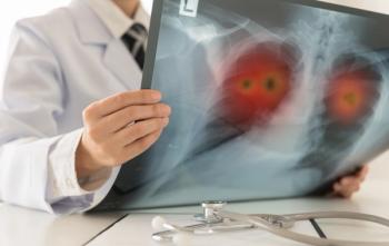
Positron emission tomography (PET) imaging has long been used to monitor the presence or progression of cancer, but a new study by a South Korean research team revealed that PET scans might also be able to detect rheumatoid arthritis (RA).
RA is an autoimmune disease which causes chronic inflammation of the joints and other areas of the body, and impacts nearly 1% of all adults.
Researchers in the
Hai-Jeon Yoon, MD, assistant professor, department of nuclear medicine at Ewha Womans University, College of Medicine, Seoul, Korea, says these proteins and the white blood cells become active as a part of the autoimmune response associated with RA and similar illnesses.
“We evaluated the value of 18F-FEDAC PET in comparison with 18F-FDG,” she says. “From our data, we found that each tracer may have different information of arthritis; 18F-FEDAC for early detection of subclinical arthritis, while 18F-FDG for measuring disease activity of symptomatic arthritis.”
These findings are expected to contribute to the improvement of personalized therapeutic outcomes by expanding the scope of molecular imaging and nuclear medicine.
“By using a 18F-FEDAC radioligand that targets TSPO, which is abundant in activated macrophages, we demonstrated that 18F-FEDAC makes it possible to see active inflammation sites in arthritic joints, and it suggests imaging with 18F-FEDAC could be useful when RA is suspected,” Yoon says. “This study is important because it showed the value of 18F-FEDAC PET as an inflammation biomarker for early detection of RA, even before onset of clinical symptoms of joints.”
Moreover, the findings suggest that early treatment can reduce the progression of joint destruction and enhance the effect of DMARDs or target drugs, because the burden of inflammatory reaction is smaller in the very early phase of RA.
“We observed that 18F-FEDAC uptake increased in paws of murine RA models in association with TSPO expression of activated macrophage, even before the onset of clinical symptoms of arthritis,” Yoon says. “18F-FEDAC can help us to find which patients will actually progress to clinically significant RA and need treatments.”
Yoon believes that the application of F18-FEDAC PET for other autoimmune diseases will be possible in the future.
“Some studies already applied similar radiotracers with F18-FEDAC, e.g. C11-PK11195 or PBR for the evaluation of systemic sclerosis or other autoimmune disease animal models,” she says.
Meanwhile, a recent Stanford University
Their study, published in Osteoarthritis and Cartilage, showed PET-MRI provides excellent, high-resolution morphologic information in patients with OA and also could serve as a way to monitor quantitative metrics relating to early biochemical changes in soft tissues affected in arthritis patients.
“Given the importance of detecting early stage OA disease in all joint tissues, there is great interest in evaluating bone remodeling as a marker of early bone degeneration and its potential as a target for disease modifying therapies,” lead author Feliks Kogan, an instructor of radiology at Stanford, wrote in the study. “As a result, there is an urgent need to develop diagnostic technologies able to quantitatively evaluate early changes in bone remodeling and its role in degenerative changes observed in adjacent tissues.”
Keith Loria is an award-winning journalist who has been writing for major newspapers and magazines for close to 20 years, on topics as diverse as sports, business, and healthcare.
Advertisement
Related Content
Advertisement


Advertisement
Advertisement
Trending on Managed Healthcare Executive
1
Payers lean on step therapy, formulary exclusions as IRA reshapes Part D benefit design
2
1,700 sites closed, 77,000 fewer children received PEPFAR-supported treatment after PEPFAR disruption | IAS 2026
3
Health Begins at Home: Why Housing Has Become a Strategic Priority for Managed Care
4
Consolidative radiotherapy shows no survival benefit, added risk in SCLC
5